Retinal Detachment
Retinal Detachment
Advanced surgical repair to reattach the retina and protect your vision
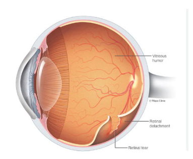
The retina is a thin layer of light-sensitive nerve cells inside the eye that allows for proper vision. Retinal detachment occurs when there are holes in the outer part of the retina, allowing fluid in the eye to seep underneath and lift the retina, similar to a bubble in wallpaper. This separation can result in bleeding and clouding of vision. For Retinal Detachment Treatment in Andheri visit Dr Vaidya Eye Hospital or Book an appointment online with Dr. Deepak Vaidya.
Most retinal detachments happen as a result of aging, but those who are nearsighted, have had cataract surgery, or have experienced a severe blow to the eye are more likely to experience it.
Some families may have a genetic predisposition for retinal detachments, but this is rare.
Treatment for a detached retina involves surgery to seal the retinal holes and reattach the retina. The surgery will be performed by an experienced eye surgeon.
Treatment Highlights
-
Cryotherapy & Scleral Buckle for sealing retinal holes
-
Vitrectomy – Keyhole surgery to repair breaks using laser or cryotherapy
-
Gas bubble, air, or silicone oil tamponade to hold the retina in place
-
Suture-less micro-incision surgical approach
-
Post-operative posturing guidance for optimal healing
-
Comprehensive follow-up within 7–14 days
Quick Facts & Details
How to perform retinal detachment surgery?
Retinal detachment surgery is usually performed under local anaesthesia, which means you will be awake during the procedure.
The eye and surrounding area will be numbed with local anaesthesia to prevent any pain. You may be aware of bright lights or movement in the operating room, but you will not be able to see details of the procedure. You will be asked to lie flat and keep your head still during the operation. General anaesthesia, where you are unconscious for the entire operation, is not commonly used for retinal detachment surgery. If you require general anaesthesia, you will need to follow specific guidelines for eating and drinking before the operation.
To correct your retinal detachment, your surgeon will perform one of the following procedures:
Cryotherapy & Scleral Buckle
Two procedures for sealing retinal holes. “Splints” or buckles made of sponge or silicone material are positioned outside the white part of the eye and under the skin, usually remaining there permanently.
Vitrectomy
A type of keyhole surgery that removes the vitreous through tiny openings. The surgeon seals the breaks in the retina using laser or cryotherapy, and a gas bubble, air or silicone oil acts as a “splint” to hold the retina in place until the tear is sealed — usually about ten days.
After a vitrectomy with a gas, air or silicone oil bubble, it is important to follow any specific instructions provided by your surgeon, including avoiding air travel, as changes in air pressure can cause the bubble to expand, leading to raised pressure in the eye and potentially causing visual loss.
Types of gas used in surgery
You will be informed of the type of gas or air bubble used in your surgery. For silicone oil, you should avoid flying for two weeks unless your consultant gives you clearance. Silicone oil may require removal during a subsequent minor procedure several months later. At the conclusion of the procedure, there may be small sutures on the eye, covered by a protective pad and clear shield. The pad will be removed the next day and you need to wear the clear shield only at night for three weeks after surgery.
It’s important to inform your doctor that you have gas in your eye when receiving medical attention, specifically during childbirth or emergency treatments, as nitrous oxide — which is frequently used for pain relief — could lead to problems in your eye if it reacts with the gas. If you require a general anaesthetic, make sure to let the anaesthetist know that you have had surgery and have gas in your eye.
Recovery After Surgery
What to expect as your eye heals — from medication to posturing — step by step.
Eye Drops
You will be given eye drops to reduce inflammation and prevent infection. Do not rub your eye, and take pain relief such as paracetamol if needed.
Healing Period
Mild discomfort and itching are normal for 5–10 days. Your eye may take 2–6 weeks to fully heal.
Follow-Up
You will have a follow-up appointment with your doctor within 7–14 days of your surgery.
Rest
Rest is important while your eye is healing. Avoid strenuous activity until your surgeon advises otherwise.
Posturing
You may need to “posture” for up to seven days — holding a set head position so the gas or oil bubble presses the retina into place.
What happens after the procedure?
After undergoing retinal detachment surgery, you will be given eye drops to reduce inflammation and prevent infection.
You should not rub your eye, and may take pain medication, such as paracetamol, if needed.
It is normal to experience discomfort and itching for 5–10 days after the surgery, and your eye may take 2–6 weeks to heal.
You will have a follow-up appointment with your doctor within 7–14 days.
It is important to rest while your eye is healing.
After your surgery, you may need to practise “posturing” for up to seven days.
This involves lying or sitting in a specific position to help the gas or silicone oil bubble in your eye float up and press the retina into position while it heals.
Your surgeon will advise you on posturing and give you instructions after your surgery.
As the gas bubble starts to disperse, you may notice a line in your vision that moves like a spirit level.
You will have clear vision above the line, but below the line your vision may be fuzzy or blurred.
The length of time the gas stays in your eye depends on the type of gas used.
After the surgery, it may take several weeks for your vision to fully recover.
If a gas bubble was used, your vision may be blurred immediately after the surgery, but this is normal.
The final outcome of your vision will depend on the state of your detached retina before the surgery.
In most cases, central vision can be restored if the retina was diagnosed and treated quickly.
However, if the affected eye already had poor central vision, full restoration may not be possible.
You may not be able to read using the affected eye, but you will still have peripheral vision which is important for daily activities such as walking and driving (if the other eye has normal vision).
When to Seek Medical Advice
Contact us promptly if you notice any of the following while your eye is healing.
Signs of Infection
Increasing redness, discharge, worsening pain, or a fever may indicate an infection that needs urgent attention.
Healing Concerns
Sudden loss of vision, a new shadow or curtain across your sight, or a marked increase in floaters or flashes of light.
Urgent Consultation
Severe or escalating eye pain that is not relieved by paracetamol, or any concern that your eye is not recovering as expected.
Why is the repair procedure so vital?
Prevents Blindness
Prompt repair stops the detachment from progressing and protects you from permanent, irreversible blindness.
Restores Vision
Reattaching the retina restores some — and often most — of the vision affected by the detachment.
Recovers Lost Sight
Timely surgery helps recover sight already lost due to the detached retina, especially when treated early.
Risks of the surgery
The success of retinal detachment surgery is not guaranteed, as each case can vary in complexity. Some patients may require multiple procedures, and your doctor will thoroughly explain the potential risks and advantages of the surgery before proceeding.
- 01 The success rate is around 85–90% with a single operation, resulting in reattachment and stability of the retina. However, there is a 5–10% chance of requiring additional surgery due to new tears, scar tissue, or redetachment.
- 02 Post-operative cataract formation is a possible side effect of the surgery, which typically requires separate treatment.
- 03 As with any surgical procedure, there is a small risk of infection and bleeding. These risks, although low, can result in permanent visual loss if they occur.
Possible complications
Complications are rare, and in most instances they can be adequately treated. Some problems may, very infrequently, result in blindness.
- 01 Bruising of the eye or eyelids can occur post-surgery.
- 02 High pressure and inflammation inside the eye are also potential side effects.
- 03 A risk of developing a cataract in the operated eye exists.
- 04 Double vision is another potential complication.
- 05 An allergy to the medication used during the procedure can occur.
- 06 Although rare, endophthalmitis (an infection in the eye) can occur, potentially leading to serious vision loss.
- 07 Cataract — People who have had a detached retina or surgery are more susceptible to developing cataracts. The treatment involves removing the cloudy lens and replacing it with a new, artificial plastic lens.
When is further surgery needed?
If you are part of the 5–10% of patients who develop another retinal tear or scar tissue, additional surgery will be required. The eye’s attempt to heal a detached retina can lead to the formation of scar tissue and retinal contraction, known as proliferative vitreoretinopathy (PVR), which is associated with decreased vision and the potential for redetachment even after successful initial surgery.
Frequently Asked Questions
Restore Clarity for Your Eyes
Experience highly advanced, personalized care for Retinal Detachment at Dr. Vaidya Eye Hospital. Book a comprehensive diagnostic exam.