Glaucoma Treatment
Glaucoma Treatment
Comprehensive Intraocular Pressure Management & Nerve Protection
What is Glaucoma?
Glaucoma is a slow, progressive eye condition that damages the optic nerve due to increased intraocular pressure (IOP). It is the world’s second leading cause of blindness and is often called the “sneak thief of sight” because vision loss may occur without early symptoms.
Early diagnosis through regular eye examinations can help prevent severe vision loss and protect long-term eye health.
Normal Vision vs Glaucoma Vision

Normal Vision
A clear, full field of view

Glaucoma Vision
Peripheral loss leading to tunnel vision
Types of Glaucoma
Glaucoma takes several forms each differs in how and why eye pressure builds up.
Chronic Open-Angle Glaucoma
Usually occurs after the age of 45 and develops gradually due to resistance in fluid outflow, raising intraocular pressure. Vision loss often starts from peripheral vision and may lead to tunnel vision.
Acute Angle Closure Glaucoma
A blockage of fluid flow causes a sudden rise in eye pressure. Symptoms include severe eye pain, headache, nausea, reduced vision, and seeing rainbow-coloured rings around lights.
Chronic Angle Closure Glaucoma
Occurs when the drainage canal of the eye is closed by the iris. It behaves almost like open-angle glaucoma.
Developmental Glaucoma
A type of angle closure glaucoma. It includes congenital glaucoma present from birth and juvenile glaucoma in children and young adults.
Normal Tension Glaucoma
A form of open-angle glaucoma where the optic nerve is highly sensitive even to normal eye pressure, gradually causing vision loss.
Secondary Glaucoma
Caused by diseases or conditions damaging the eye drainage system including diabetes, arthritis, cataract, eye injuries, inflammation, long-term steroid use and eye tumours.
Causes of Glaucoma
Our eyes constantly produce a clear fluid called aqueous humour. It flows through the pupil and behind the cornea, then drains through the trabecular meshwork. In glaucoma, the fluid does not drain freely, increasing intraocular pressure.
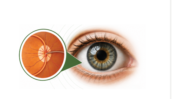
Raised intraocular pressure damages the optic disc and the nerve fibres responsible for vision. Glaucoma usually affects peripheral vision and may cause tunnel vision in later stages.
Fluid Production
Aqueous humour is made
Drainage Issue
Fluid cannot drain freely
Increased Eye Pressure
Intraocular pressure rises
Optic Nerve Damage
Nerve fibres are harmed
Vision Loss
Peripheral / tunnel vision
Who is at Risk?
Age above 40
Risk rises with age
Family History
A relative with glaucoma
Long-Term Steroid Use
Prolonged steroid medication
Coloured Rings Around Lights
A warning visual symptom
Diabetes or Hypertension
Systemic health conditions
Eye Injuries
Past trauma to the eye
Tests Performed to Diagnose Glaucoma
Tonometry
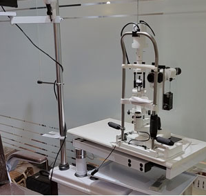
Measures pressure in the eye using an Applanation Tonometer attached to a slit lamp (a handheld version also exists). Non-contact air-puff instruments are good for screening but cannot diagnose glaucoma. Average eye pressure is about 18 mm Hg higher-than-average pressure doesn't always mean glaucoma.
Ophthalmoscopy
Medicated drops dilate the pupil to examine the optic disc and the back of the eye, the retina. A 72-D lens gives a stereoscopic view, and a computerised scan of the optic disc can also be done.
Optical Coherence Tomography (OCT)
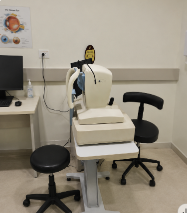
Very useful for a glaucoma suspect or early-to-moderate disease. It detects damage and progression of optic nerve fibres and gives structural information about the retina, optic nerve head and lamina cribrosa. Newer OCT angiography (OCTA) maps retinal blood vessels and may help in advanced stages.
Gonioscopy
After numbing the eye, the angle of the eye is examined with a gonioscope a special lens gently placed on the surface of the eye. It allows a more accurate diagnosis of the type of glaucoma.
Pachymetry
Measures the thickness of the cornea. Corneal thickness can increase effort in reading and similar activities, and also increases the risk of having glaucoma.
Perimetry
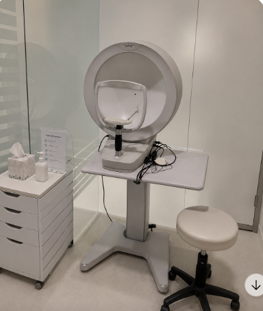
Automated perimetry confirms damage to the optic nerve. In early stages, glaucoma can only be detected or monitored this way. A person with glaucoma has areas missing from their field of vision. Periodic examinations are essential to check progression.
Controlling Glaucoma
Glaucoma can be controlled with treatment, but it cannot be cured. The aim is to lower eye pressure and prevent further optic nerve damage and vision loss.
Treatment with Eye Drops
Medicines lower eye pressure. Many glaucoma patients need lifelong medicines to control the condition.
Tablets
Acetazolamide / Diamox tablets may be given to reduce aqueous humour production inside the eye.
Laser Treatment
Argon Laser Trabeculoplasty (ALT)
Commonly used for open-angle glaucoma. The laser treats the trabecular meshwork to increase drainage outflow and lower IOP though medication is often still needed. Usually half the meshwork is treated first and the rest later, reducing the risk of a pressure spike. ALT has successfully lowered eye pressure in up to 75% of patients and can be performed two to three times per eye over a lifetime.
Laser Peripheral Iridotomy (LPI)
Used for angle-closure glaucoma. It makes an opening through the iris, allowing aqueous fluid to flow from behind the iris directly to the front chamber, bypassing its normal route. LPI is the preferred method for a wide variety of angle-closure glaucoma and is most often used to treat an anatomically narrow angle and prevent attacks.
Cyclophotocoagulation & ECP
Two laser procedures that reduce aqueous humour by destroying part of the ciliary body usually reserved for eyes with elevated IOP after other treatments fail, or where filtering surgery isn't possible. Transscleral cyclophotocoagulation directs energy through the sclera; with ECP, the instrument is placed inside the eye so laser energy is applied directly to the ciliary body.
Incisional Glaucoma Surgery
Glaucoma surgery is performed as an outpatient procedure, usually with local anaesthesia (with or without intravenous sedation) under an operating microscope. Afterwards, patients can wear their glasses and usually wear a protective shield only at bedtime for one to two weeks.
The most common incisional surgery, trabeculectomy, creates an alternative channel to divert aqueous fluid from the eye and usually takes about 25–35 minutes. It often reduces or eliminates the need for future glaucoma medications. For patients more at risk of scarring, the surgeon may use an anti-fibrosing medication such as 5-fluorouracil or mitomycin C.
Trabeculectomy at a Glance
Takes about 25–35 minutes
Local anaesthesia, outpatient procedure
Creates a new channel to drain fluid
Often reduces future medication needs
Can a Person Become Blind from Glaucoma?
This depends on the stage at which glaucoma is diagnosed. Early diagnosis reduces the chances of blindness. Glaucoma can often be stabilised with medicines or surgery — which is why regular eye check-ups are so important.
Restore Clarity for Your Eyes
Experience highly advanced, personalized care for Glaucoma Treatment at Dr. Vaidya Eye Hospital. Book a comprehensive diagnostic exam.